General Information
Strain Name | NOD.Cg-Tg(tetO-Alb-uPA)1Deng Rag2tm1Il2rgtm1/Vst |
Common Name | URG®;Tet-uPATgRag2null Il2rgnull |
Origin | Beijing Vitalstar Biotechnology Co., Ltd. |
Background | NOD |
Coat color | Albino |
Development
URG mice were obtained by crossing four genetically modified animal models containing two transgenes and two knockouts. The constructs were performed as follows:
First, TRE-uPA transgenic mice were crossed with Alb-rtTA transgenic mice to obtain Alb-rtTA/TRE-uPA double-positive mice, in which uPA expression and liver injury were detected in the livers by Dox induction [1]; second, Alb-rtTA/TRE-uPA double-positive mice were backcrossed for 8 consecutive generations to NRG (NOD background of Rag2nullIl2rgnull mice) background to obtain Alb-rtTA/TRE-uPA/Rag2null/Il2rgnull mice (URG). uPA was not expressed when URG was not induced, and the mice grew healthily; when induced with doxycycline (Dox), uPA driven by the Tet-On system was then expressed in the liver, and the mice developed liver injury. expression, and the mice developed liver injury.
Phenotype
1. Time to liver injury can be regulated
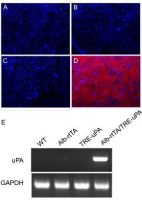
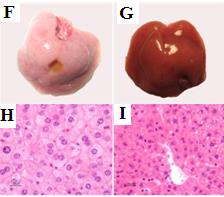
Fig 1. Analysis of liver injury in URG® mice
URG® are routinely expanded like NPG mice and can be induced with Dox at any time to induce liver injury. In the absence of induction, expression of uPA expression was undetectable (C); after Dox induction, uPA expression was detectable by immunohistochemistry (D); uPA-expressing livers were pale (F); livers of wild-type mice were dark red (G); hepatocyte swelling and vacuolization were seen in HE staining of URG® mice (H); hepatocyte morphology was normal in control mice (I).
2. URG® Serological Indicators after Liver Injury[2]
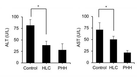
Figure 2. serum levels of ALT and AST in URG® mice induced by Dox drinking water (n=5/group)
Note: Hepatocyte-like cells (HLC), primary human hepatocytes (PHHs), and PBS were used as controls and transplanted into mice via intrasplenic transplantation, respectively. Serum ALT and AST levels were determined 7 weeks after transplantation
Examples of URG® mouse studies
1. Validation of the regenerative capacity of functional hepatocytes (EPS-Heps) in vivo[3]
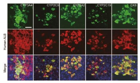
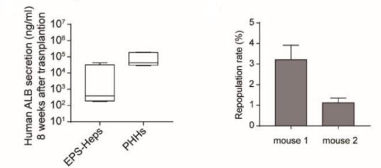
Fig 3. Human Hepatocyte Reconstruction in URG® Mice Transplanted with EPS-Heps vs. PHHs 8 Weeks Later
Eight weeks after transplantation of EPS-Heps in URG® mice, co-expression of human CYP3A4, CYP2C9, CYP1A2, and CYP2C19 with human Alb was detected, as well as secretion of human Alb in serum. These data suggest that EPS-Heps can reconstruct damaged mouse livers characterized by functional maturation.
2. For hepatocyte-like cell (HLC) transplantation in the treatment of liver failure[2]
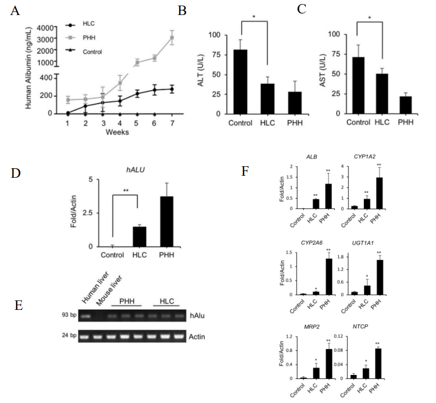
Fig 4. Hepatocyte regeneration after transplantation of HLC in URG® mice
8-week-old male mice (n=5/group) were injected with 15 mg/kg body weight of Dox via intraperitoneal injection 12 hours prior to cell transplantation, and HLCs, PHHs, were transplanted intrasplenically into the mice using PBS as a control. After cell transplantation URG® mice were continuously fed Dox-containing drinking water for 6 weeks. Blood was taken from the tail vein every week, and human ALB levels were quantified by ELISA, and the mice were put to death at week 7. The results showed that URG® mice transplanted with HLC showed a gradual increase in serum human ALB secretion and a significant decrease in serum ALT and AST concentrations after 7 weeks. Real-time PCR of human-specific ALU sequences further confirmed the colonization of HLC in the mouse liver. Human-specific gene ALB and human hepatocyte-specific metabolic genes phase I enzymes CYP1A2 and CYP2A6 were also detected. These data suggest that HLC can be integrated into the liver of URG® mice to ameliorate liver dysfunction.
3. In vivo validation test of the function of transdifferentiated liver cells in vitro[4]
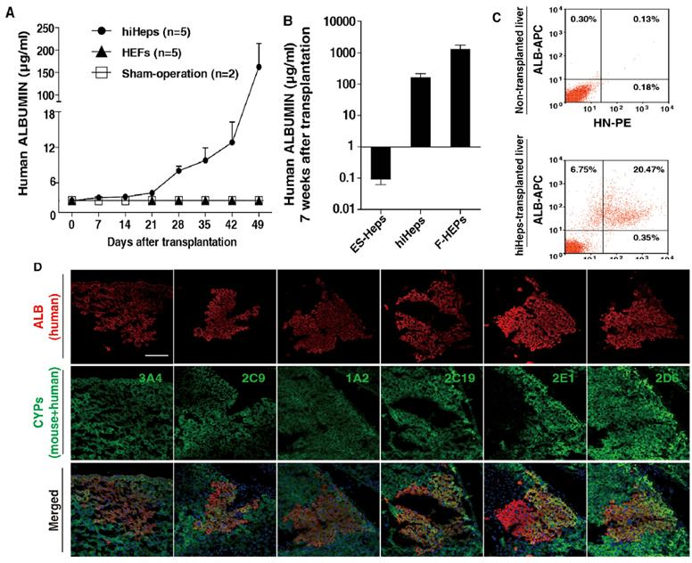
Fig5. Hepatocyte regeneration after transplantation of human induced hepatocytes (hiHeps) in URG® mice
URG® mice were injected intra-splenically with ES-Heps (n=16), hiHeps (n=5) and F-HEPs (n=6). Blood samples were collected, human Alb was quantified by ELISA, and the liver sections were stained and flow analyzed. The results showed that human Alb secretion in the serum of mice gradually increased after transplantation of hiHeps in URG® mice, and clusters of human Alb cells were observed to be expressed in mice 6 weeks after transplantation. The metabolic function of hiHeps in vivo was also confirmed, and the expression of CYP3A4 and CYP2C9 was analyzed. It indicates that hiHeps can grow stably and functionally in vivo. This study illustrates that URG® mice can be used to validate hiHeps as a potential cell source for hepatocyte transplantation.
URG® Mice Applications
1. Modulated liver injury and humanized liver injury models can be constructed for human-specific liver disease, drug safety assessment and human-specific liver metabolism enzyme-related research
2. HBV, HCV infection and antiviral activity signaling pathway research
3. Drug metabolism and pharmacokinetics, drug efficacy studies
4. Stem cell research
5. Gene therapy
References
1. Song X J, et al. A Mouse Model of Inducible Liver Injury Caused by Tet-On Regulated Urokinase for Studies of Hepatocyte Transplantation. Am J Pathol. 2009, 175(5):1975-1983.
2. Li Z, et al. Generation of qualified clinical-grade functional hepatocytes from human embryonic stem cells in chemically defined conditions. Cell Death Dis. 2019, 10:763.
3. Wang Q, et al. Generation of human hepatocytes from extended pluripotent stem cells. Cell Res. 2020, 30:810-813.
4. Du Y, et al. Human hepatocytes with drug metabolic function induced from fibroblasts by lineage reprogramming. Cell Stem Cell. 2014, 14(3):394-403.

 animalmodel@vital-bj.com
 +8610-84928167