General Information
Strain Name | C57BL/6N -Tg(1.28mer HBV)6 /Vst |
Common Name | C57BL/6-HBV; HBV-Tg mice |
Origin | Beijing Vitalstar Biotechnology Co., Ltd. |
Background | C57BL/6NCrl |
Coat color | Black |
Development
A 1.28-fold linearized fragment of HBV (type A, GenBank: AF305422.1) was injected into the embryonic nucleus of C57BL/6NCrl mice to obtain transgenic positive mice. The first mice with a copy number of 107-108 IU/mL were retained by peripheral blood HBV copy number analysis. An HBV transgenic mouse line was established according to the hemizygous pairing with wild C57BL/6NCrl mice.
This mouse produces intact, infectious viral particles in vivo, and the level of HBV replication is comparable to that of a chronic hepatitis B patient. In addition to high titers of HBV DNA, high levels of HBsAg and HBeAg were detected in peripheral blood.

Fig 1. Schematic diagram of the HBV-Tg mouse strategy
Genotype identification information

Fig 2. Genotyping of HBV-Tg mice
Phenotype
1. Real-time quantitative PCR of HBV DNA, HBeAg and HBsAg in peripheral blood
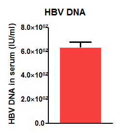
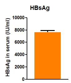
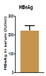
Fig 3. HBV DNA, HBeAg and HBsAg levels in peripheral blood of HBV-Tg mice
2. Results of HBV virological assay in liver tissues of HBV-Tg mice at different weekly ages[1]

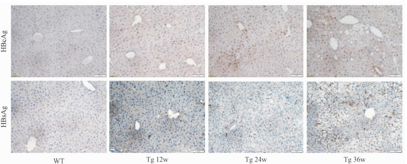
Figure 4. Serum HBsAg, HBeAg, HBV DNA levels and liver tissue HBcAg and HBsAg expression (×200)
Note: WT is 36-week-old wild-type male mice, Tg 12 w is 12-week-old HBV-Tg mice, Tg 24 w is 24-week-old HBV Tg mice, and Tg 36 w is 36-week-old HBV Tg mice
The results showed a stable HBV virological profile by observing HBV-Tg mice at different weekly ages.
3. Liver function tests and HE staining in HBV-Tg mice at different weekly ages[1]

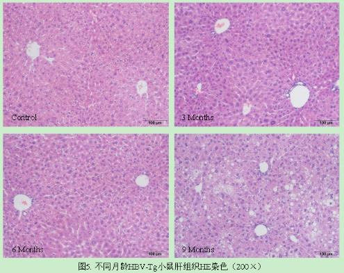
Fig5. HE staining of liver tissue of HBV-Tg mice at different weekly ages (×200)
The results showed that HE staining showed that the liver lobules of wild-type mice were structurally intact and clear, and the hepatocyte cords were neatly arranged and radially distributed around the central vein; the liver tissues of HBV-Tg mice had enlarged nuclei at 12 and 24 weeks of age, and showed obvious pathological changes such as steatosis, ballooning, and disorganized arrangement of hepatocyte cords at 36 weeks of age.
4. Sirius red staining of liver tissues of HBV-Tg mice at different weekly ages[1]
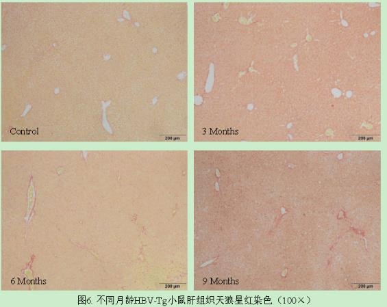
Figure 6. Sirius red staining (×100) of liver tissues of HBV-Tg mice at different weekly ages
The results showed that the liver tissues of 6- and 9-month-old HBV mice showed a small amount of collagen deposition, which was mainly concentrated in the confluent area and between the hepatic lobules, and bridging and pseudolobules were not seen to appear.
HBV-Tg Mice Applications
1. Study on the anti-HBV activity of polysaccharide substances[2]
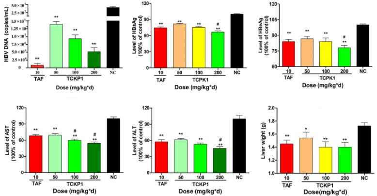

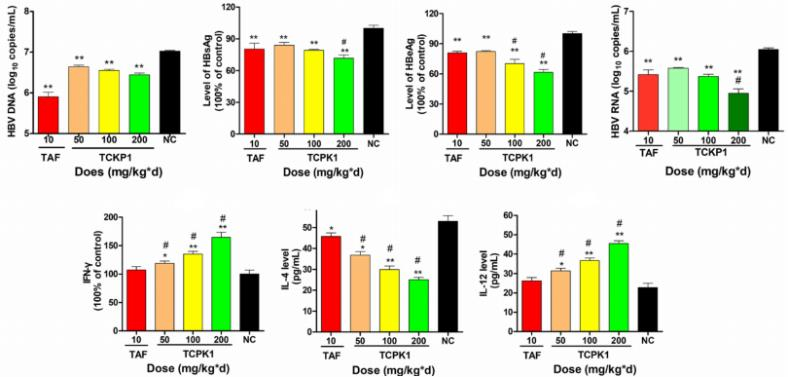
A B C
Fig7. Anti-HBV activity of TCKP1
Twenty-five 5-week-old SPF-grade HBV-Tg mice were randomly divided into five groups and injected intraperitoneally daily with 10 mg/kg-d TAF (positive control group), TCKP1 low-dose, medium-dose, and high-dose groups as well as negative control group (PBS). Blood was taken 28 days after administration, and livers were analyzed after mice were executed. The results showed that TCKP1 significantly reduced serum HBsAg, HBeAg, HBV DNA, ALT, AST levels, as well as HBsAg, HBeAg, HBV DNA and HBV RNA levels in the livers, and liver congestion and swelling were improved to different degrees when compared with the negative control group. Elevated serum IL-12 and IFN-γ levels were also observed. These results suggest that the anti-HBV effect of TCKP1 is realized by enhancing the function of immune cells and regulating the balance of Th1/Th2 cytokines.
2. Studies on the regulation of HBV efflux regulators[3]

A B
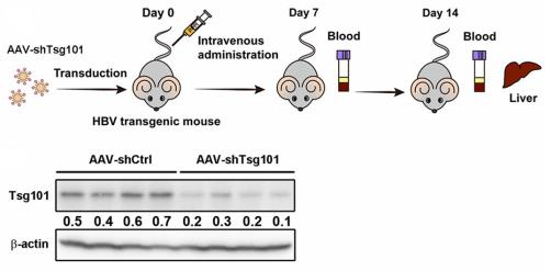
Fig8. Effect of knockdown of TSG101 on the secretion of HBV viral particles
Eight 6-8-week-old HBV-Tg mice were randomized into two groups. Recombinant AAV (1×10¹² viral genome) targeting shRNA or Tsg101 was injected via tail vein. Serum HBsAg and HBV DNA levels were determined 7 d and 14 d after the first administration. The level of Tsg101 in the liver was determined 14 days after the first administration. The results showed that knockdown of TSG101 significantly inhibited serum HBsAg and HBV DNA in HBV-Tg mice, confirming that TSG101 recognizes ubiquitinated HBc proteins and enables HBV to utilize the MVB pathway to exit the cell, which provides a new target for antiviral drug development.
3. Evaluation of new anti-HBV drugs
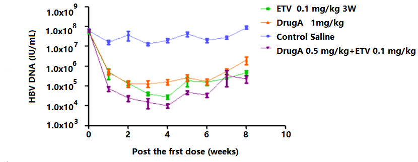
Fig. 9. Evaluation of efficacy of different drugs, doses and combinations in HBV-Tg mice
4. CRISPR-Cas9 Technology for HBV[4]
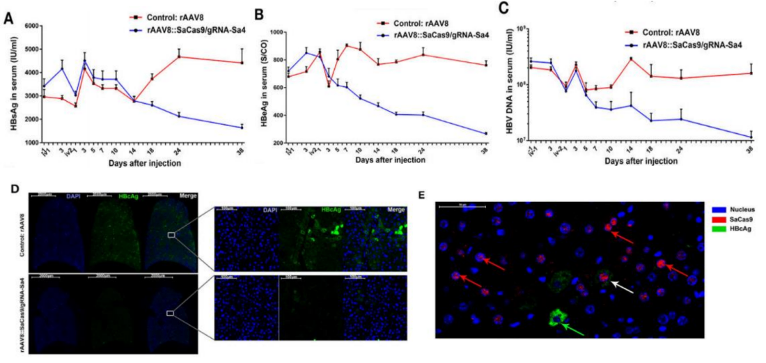
Fig 10. Inhibition of HBV antigen expression and HBV replication in HBV-Tg mice by CRISPR-SaCas9
Fourteen HBV-Tg mice were randomly divided into test and control groups (n=7/group), and blood was drawn from the eye vein of the mice on the 3rd day after the first administration, and the 1st, 3rd, 5th, 7th, 10th, 14th, 18th, 24th, 38th, and 53rd days after the second administration. On the 39th day after the second injection, three mice were randomly selected from each group and dissected for heart, liver, spleen, lungs and kidneys, and tissue sections were prepared for observation. The results showed that the levels of HBV markers in the serum of mice in the test group continued to decrease over time, and the fluorescence intensity of HBcAg staining of liver tissues was lower, indicating that the expression of HBcAg in the hepatocytes of mice could be effectively inhibited.
HBV-Tg Mice Applications
1. the model can be used to study HBV, therapeutic drugs and vaccines and further downstream in chronic hepatitis and fibrosis (combined with chemical induction).
2. It provides a genetically stable model for studying viral and host factors affecting HBV replication, pathogenetic and physiological processes
References
孙鑫,等. C57BL/6N-Tg(1.28HBV)/Vst 乙肝病毒转基因小鼠肝脏炎症与纤维化的病理特点. 肝脏, 2018, 23(01):26-30.
Tang F, et al. Anti-HBV Activities of Polysaccharides from Thais clavigera (Küster) by In Vitro and In Vivo Study. Mar Drugs. 2021, 19(4):195.
Zheng Y, et al. Hepatitis B virus hijacks TSG101 to facilitate egress via multiple vesicle bodies. PLoS Pathog. 2023, 19(5):e1011382.
Li H, et al. Inhibition of HBV Expression in HBV Transgenic Mice Using AAV-Delivered CRISPR-SaCas9. Front Immunol. 2018, 9:2080.

 animalmodel@vital-bj.com
 +8610-84928167