General Information
Origin | Beijing Vitalstar Biotechnology Co., Ltd. |
Background | HBV-Tg mice |
Coat color | Black |
Modeling methodology
A mouse composite model of hepatitis B liver fibrosis can be successfully established by intraperitoneal injection of CCl4 into HBV-Tg mice. Using 0.5-0.7 μL/g body weight of pure CCl4 (dissolved into olive oil, corn oil, or paraffin oil), intraperitoneal injections were given 2-3 times per week for 4-8 weeks.HBV-Tg mice were able to stabilize the model after 8-12 weeks of combined CCl4 injections.
Due to the strong regenerative ability of the mouse liver and the good tolerance of the mouse liver to CCL4, the ALT and AST generally recovered or approached normal levels after the metabolism of CCL4 was completed about 3 days after the injection. Therefore, the therapeutic experiments of CCL4 or combined with HFD need to take into account the characteristics of the changes in liver injury indexes, as well as the details of the setting and sampling analysis of the control and treatment groups.
Phenotype
1. Changes in ALT and AST levels in peripheral blood of mice after CCL4 injection
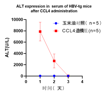
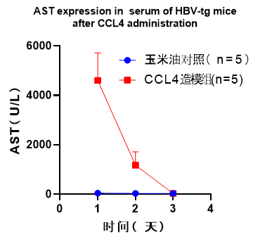
Fig 1. serum ALT and AST levels induced by CCL4 in HBV-Tg mice
2. Characteristics of changes in ALT and AST levels in peripheral blood at 12 to 16 weeks
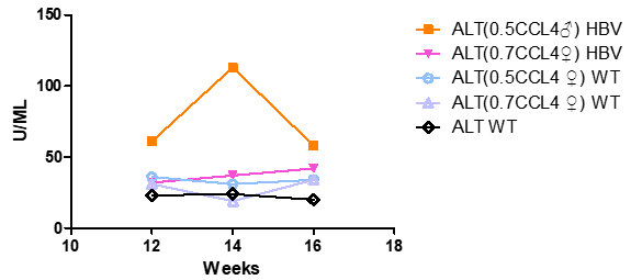
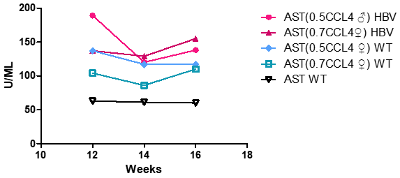
Fig 2. serum ALT and AST levels induced by CCL4 in HBV-Tg mice
HBV-Tg mice were more sensitive than wild C57 mice, with higher peripheral blood levels of ALT and AST after the same CCL4 induction.
3. HE Staining with Sirius Red Staining
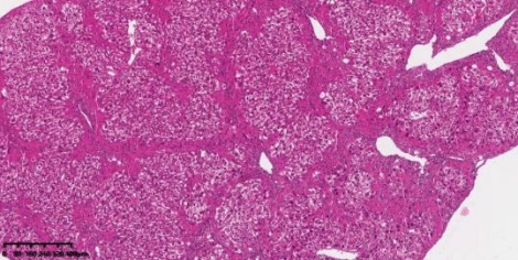
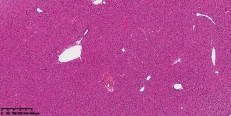
Fig 3. HE staining of liver tissues of HBV-Tg mice after 8 weeks of CCL4 induction, with the CCL4-induced group on the left and the control group on the right (injected with corn oil)
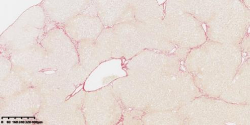
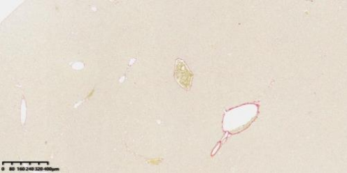
Fig 4. Sirius staining picture of liver tissue after 8 weeks of CCL4 induction in HBV-Tg mice
Note: The left side is the CCL4 induction group, and the right side is the control group (injected with corn oil). 8 to 10 weeks of CCL4 induction can result in 100% fibrosis and cirrhosis features (scores of 3 and above, with bridging features appearing on Sirius scarlet staining)
Example of HBV-Tg mice + CCL4 study
1. CCL4 induction in HBV-Tg mice can be used in liver fibrosis models[1]

Fig 5. Serum Hepatitis B Virology in Mice
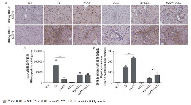
Fig 6. Immunohistochemical staining and semi-quantitative analysis of HBsAg and HBeAg in mouse liver tissue

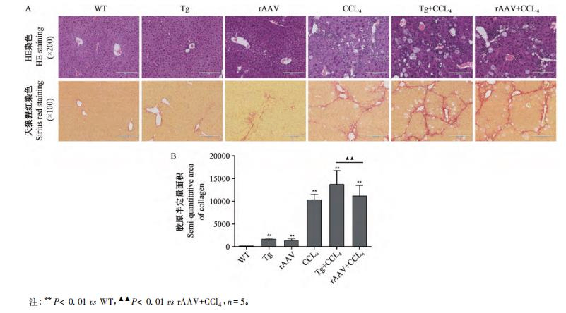
Fig 7. Liver histopathology HE, Wolf Scarlet staining with semi-quantitative analysis of collagen
C57BL/6N wild-type mice were randomly divided into wild-type control group (WT), rAAV8-1.3 HBV transfected group (rAAV), CCL4 group (CCL4), and rAAV8-1.3 HBV-transfected composite CCl4 group (rAAV+CCL4), and the same-week-old HBV-Tg mice were randomly divided into HBV-transgenic group (Tg), HBV-transgenic compound CCl4 group (Tg+CCL4). Each group consisted of 5 mice. rAAV8-1.3 HBV transfection was performed by tail vein injection, and each mouse was injected once on the first day of the experiment with a viral load of 1×10 v.g. CCL4 was diluted to 10% in olive oil and injected intraperitoneally into 2 mL/kg of mouse body weight once every other day. rAAV8-1.3 HBV transfections were performed by anesthesia at the end of the first day of the experiment, and the serum and liver tissues were retained after 12 weeks of the anesthesia and the mice were put to death. The results showed that HBsAg and HBeAg levels were significantly increased in the Tg+CCL4 group, and the liver tissues were positive for HBsAg and HBcAg. Serum ALT, AST and liver tissue Hyp levels were significantly increased in the Tg+CCL4 group, as well as the inflammatory reaction and collagen deposition. In conclusion, fibrosis progressed significantly in HBV-Tg mice.
HBV-Tg Mice+CCL4 Applications
1. Provide a stable and reliable small animal model for the study of the influence of viral and host factors on HBV replication and disease pathology.
2. To study the pharmacological efficacy of anti-HBV therapeutic drugs, including replication inhibitors, immunotherapy and gene therapy.
3. Research on the treatment of liver fibrosis
References
1. 黄恺, 孙鑫, 赵志敏, 等. 两种乙肝肝纤维化小鼠复合模型的比较. 中国实验动物学报. 2019, 27(5):598-603.

 animalmodel@vital-bj.com
 +8610-84928167