Liver cancer refers to malignant tumors that occur in the liver or start from the liver. According to GLOBOCAN statistics for the year 2023, globally, liver cancer is the fourth leading cause of cancer-related deaths, following lung cancer, breast cancer, and colorectal cancer. By studying the mechanisms of liver cancer occurrence, its development process, and pathological characteristics, we can gain a deeper understanding of the risk factors that lead to liver cancer, such as viral infections (such as hepatitis B and hepatitis C), chronic liver disease, cirrhosis, as well as environmental and genetic factors. Through mouse models, researchers can simulate the pathological process of human liver cancer, explore the role of oncogenes, screen for potential drugs, and assess the efficacy and safety of new treatment methods.
Vitalstar currently offers liver cancer models and pharmacological and efficacy services based on these models. The liver cancer models provided can be roughly divided into:
1. Syngeneic/xenograft liver cancer animal models, such as transplanting various liver cancer CDX or PDX into highly immunodeficient mice like NPG, or mouse liver tumor cell lines or tissue blocks into the same background mice, commonly used for screening new anticancer drugs and preclinical efficacy study.
2. Induced animal liver cancer models, such as DEN+CCL4 induced liver cancers in C57BL/6 mice, often used for research on liver cancer etiology, pathogenesis, pathological mechanisms, and genetics.
3. Transgene and induction combined liver cancer models, such as HBV transgenic mice induced by CCL4, Ldlr KO mice fed a WD diet, etc., commonly used to study the function of specific genes and the interaction of different genes in the development of hepatocellular carcinoma.
4. Spontaneous liver cancer models, such as KPA spontaneous liver cancer mice.
Model Name | Species | Model Feature | |||||
Body Weight | Fasting Glucose | Steatosis | Hepatitis | Fibrosis | Hepatocellular Carcinoma | ||
HFD (B6/SD/MC4R KO) | Rat and mouse | Increase | Elevate | 8-16W for mice, 8-24W for rats | 12-24W | Mild | No |
GAN NASH | Rat and mouse | Increase | Elevate | 10-12W | 12-16W | ≥12M, moderate | No |
MCD | Rat and mouse | Decrease | Reduce | 4W, mild | 4W, mild | 4-6W | No |
CDAHFD | Mouse | Invariant | Increase | 8-12W | 8-12W | 8-12W | 24-36W, 100% |
HBV-Tg=CCl4 | Mouse | Increase | Invariant | No | 4-6W | 6-8W | 24W |
HFD+CCl4 | Mouse | Increase | Elevate | 8-12W | 8-12W | 16W, F3 | 24W |
Alb/Pten KO | Mouse | Invariant | Invariant | No | No | 10-40W | 70-78W, 100% |
Alms1 KO+WD | Mouse | Increase | Elevate | 4W | 4W | 12W, F3 | 24W, 75% |
Ldlr KO+WD | Mouse | Increase | Elevate | 8-12W | 8-12W | 8-12W | 24W, 100% |
Hu-URG+CDAHFD | Humanized liver mouse | Invariant or slightly decrease | Elevate | 4-8W | 4-8W | 4-12W | 20-24W |
(1)Syngeneic/Xenogeneic Transplantation Liver Cancer Animal Models
According to the source of cell lines, the tumor engraftment mouse models can be divided into syngeneic (mouse-derived tumor cells) and xenogeneic (human-derived tumor cells) models. According to the transplantation site, the models can be further divided into subcutaneous implanted and orthotopic implanted models. Tumor mouse models can select immunodeficient mice such as nude mice, NOD-scid, and NOD/SCID/IL2Rγ(null) mice, as well as humanized immune system mice (Hu-HSC-NPG and Hu-PBMC-NPG).
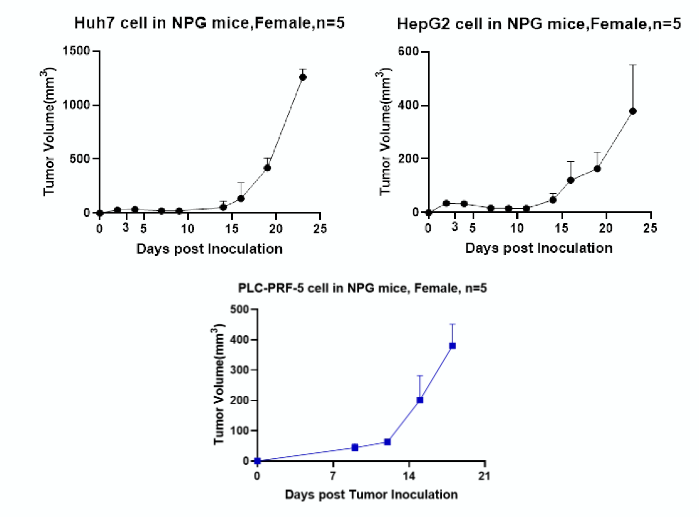
The liver cancer cell lines available at Vitalstar include: HepG2, SK-Hep1, Hep3b, SMMC-7721, SNU423, HuH7, PLC-PRF-5(HBV), SNU182(HBV), HepAD38(HBV), HepG2.2.15(HBV), H22 (mouse-derived).

Figure 1. Growth curves of tumors seeded with different human cancer liver cell lines
(2)Induced Liver Cancer Models
Many chemical carcinogens can induce tumor formation in animals when administered at sufficient doses and durations. Commonly used hepatocarcinogens include diethylnitrosamine (DEN), aflatoxin B1 (AFB1), and carbon tetrachloride (CCL4), among others.
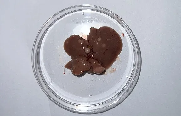
The figure below demonstrates the tail vein injection of 30 mg/kg DEN into C57BL/6 mice at 12 days of age. Starting from 4 weeks of age, the mice are given a tail vein injection of 2 uL/g of 25% CCL4 (dissolved in olive oil) once a week for 30 weeks to induce liver cancer.

Figure 2. Liver tumors induced by DEN+CCL4
(3)Spontaneous Liver Cancer Models
Cross Alb-CreERT2 mice with LSL-KrasG12D/+ and LSL-Trp53R172H/+ mice to obtain LSL-KrasG12D/+;LSL-Trp53R172H/+;Alb-CreERT2 mice. Cross Alb-CreERT2 mice with LSL-KrasG12D/+ and Pten flox mice to obtain LSL-KrasG12D/+;Pten flox;Alb-CreERT2 mice. Activating mutation of Kras, and simultaneous loss of Tp53 or Pten in hepatocytes can drive cholangiocellular and hepatocyte-derived cholangiocarcinoma occurring. After 6 days of tamoxifen induction, “Stop” signal sequences being excised from transgenic cassettes, results mouse hepatocytes undergoing expression of activating Kras and Pten/P53 deletion, even heterozygous deletion, causing ICC and HCC.

 animalmodel@vital-bj.com
 +8610-84928167