Vitalstar In Vivo Imaging Platform is equipped with the Lumina III in vivo imaging system from PE IVIS Spectrum, capable of detecting and imaging bioluminescence catalyzed by luciferase and fluorescence. We provide services for in vivo bioluminescence and fluorescence detection and imaging analysis for cells, mice, and rats.
Application Scenarios:
1. In vitro detection of luminescence intensity of cells expressing luciferase or fluorescent proteins.
2. In vivo tracking, imaging, and luminescence intensity detection of cells labeled with luciferase, fluorescent proteins, or fluorescent dyes.
3. In vivo imaging analysis of orthotopic tumors or tumor metastasis.
4. Imaging analysis of the distribution of fluorescent probes in vivo (both in live subjects and in dissected organs).
5. Imaging analysis of the expression level and in vivo distribution of gene therapy vectors labeled with fluorescent proteins or luciferase (both in live subjects and in dissected organs).
6. Imaging analysis of the in vivo distribution of drugs labeled with fluorescence (both in live subjects and in dissected organs).
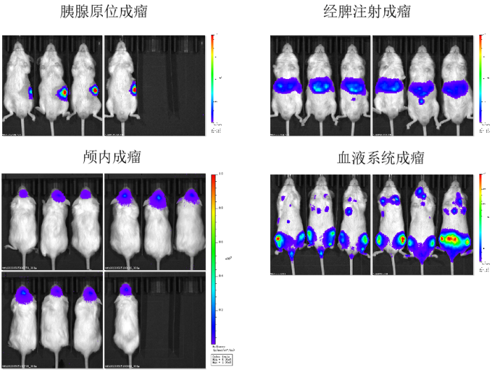
Case I: Monitoring of tumor formation in NPG mice

Figure1. Monitoring of tumor formation in pancreas, liver (via spleen injection), intracranial and blood (via tail vein injection) of NPG mice with IVIS
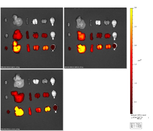
Case II: Detection of the distribution of drug labeled with fluorescence dye

Figure 2. Detection of fluorescence-labeled drug distribution in dissected mouse orgons

 animalmodel@vital-bj.com
 +8610-84928167